28 Jul Video-based pupillometry using Fourier Mellin image correlation
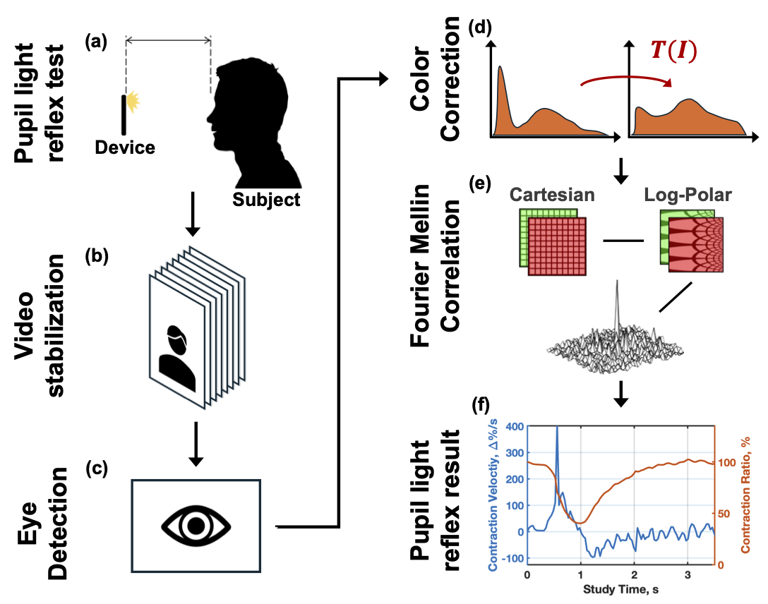
We introduce a novel method for evaluating the pupil light reflex (PLR) response using digital video recordings. Expensive, specialized devices are replacing traditional penlight tests in emergency and neurotrauma departments, but they are not widely accessible elsewhere. Recently, smartphone applications have emerged as alternatives, but...


